


Diagnosing blocked tubes is commonly done with an HSG
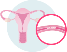 A minimally invasive fertility test called an HSG, or hysterosalpingogram, allows your TFC physician to evaluate both your uterus and your fallopian tubes. During the HSG, dye is carefully injected into your cervix using a thin catheter. Low dose XRay is used to watch the dye as it flows through the uterus and into the fallopian tubes. The dye will then either flow freely through the tube and exit from the end near the ovary, or the dye will stop flowing – indicating a blockage.
There are three sites along the tube where blockages most commonly occur.
- Obstruction at the cornual portion of the tube. This is where the tube connects to the uterus and is referred to as “proximal occlusion”
- Obstruction at the “distal” end of the tube. where the finger-like projections (“fimbia”) responsible for picking up eggs when they ovulate are located
- Least commonly, obstruction at the mid portion of the tube. This almost always results from a previous tubal ligation procedure.
If we see one blocked fallopian tube, we are suspicious that the other tube may be damaged as well, even if X-ray films do not show an obstruction. While it is common for the HSG to show damage to only one tube, at the time of corrective surgery, damage is frequently noted in the other tube as well.
Sometimes the fallopian tubes may appear to be patent, or open, when they are actually damaged and unable to function properly.